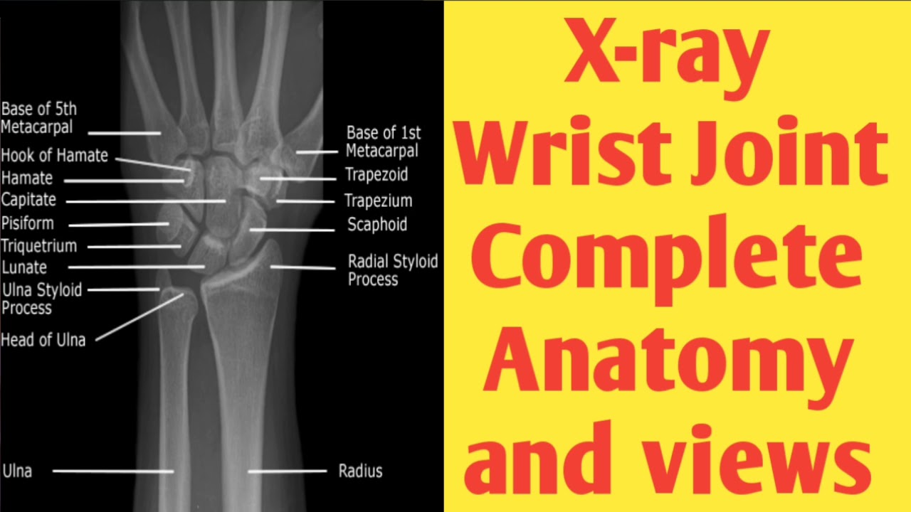
Xray wrist joint complete anatomy and views YouTube
A wrist X-ray (radiograph) is a test that produces an image of the inside of your wrist. The image displays the inner structure ( anatomy) of your wrist in black and white. A wrist X-ray shows your two forearm bones (radius and ulna) and eight wrist bones (carpal bones).

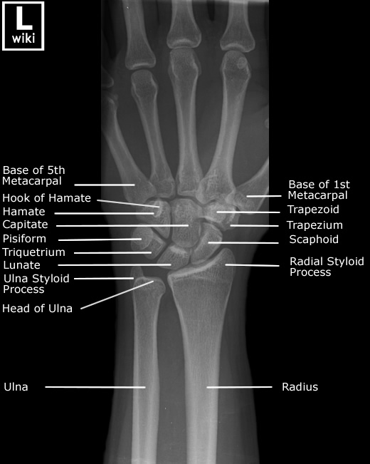
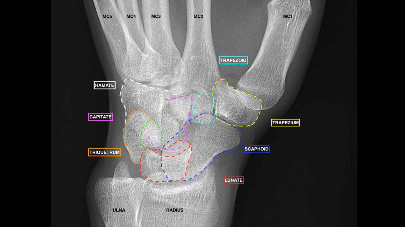
[Figure, Wrist xray with labeled osseous anatomy] StatPearls NCBI Bookshelf
Standard projections for the wrist are Postero anterior and lateral views. Some authors also consider oblique view as part of the standard views. [ 6] The following section of this article describes the standard radiographic views first and proceeds to describe any ancillary views of the same and finally dynamic studies using these projections.
Wrist Joint Anatomy Concise Medical Knowledge
The wrist joint is formed by an articulation between: Distal end of the radius and the articular disk. Proximal row of the carpal bones (except the pisiform). Together, the carpal bones form a convex surface, which fits into the concave shape of the radius and articular disk.
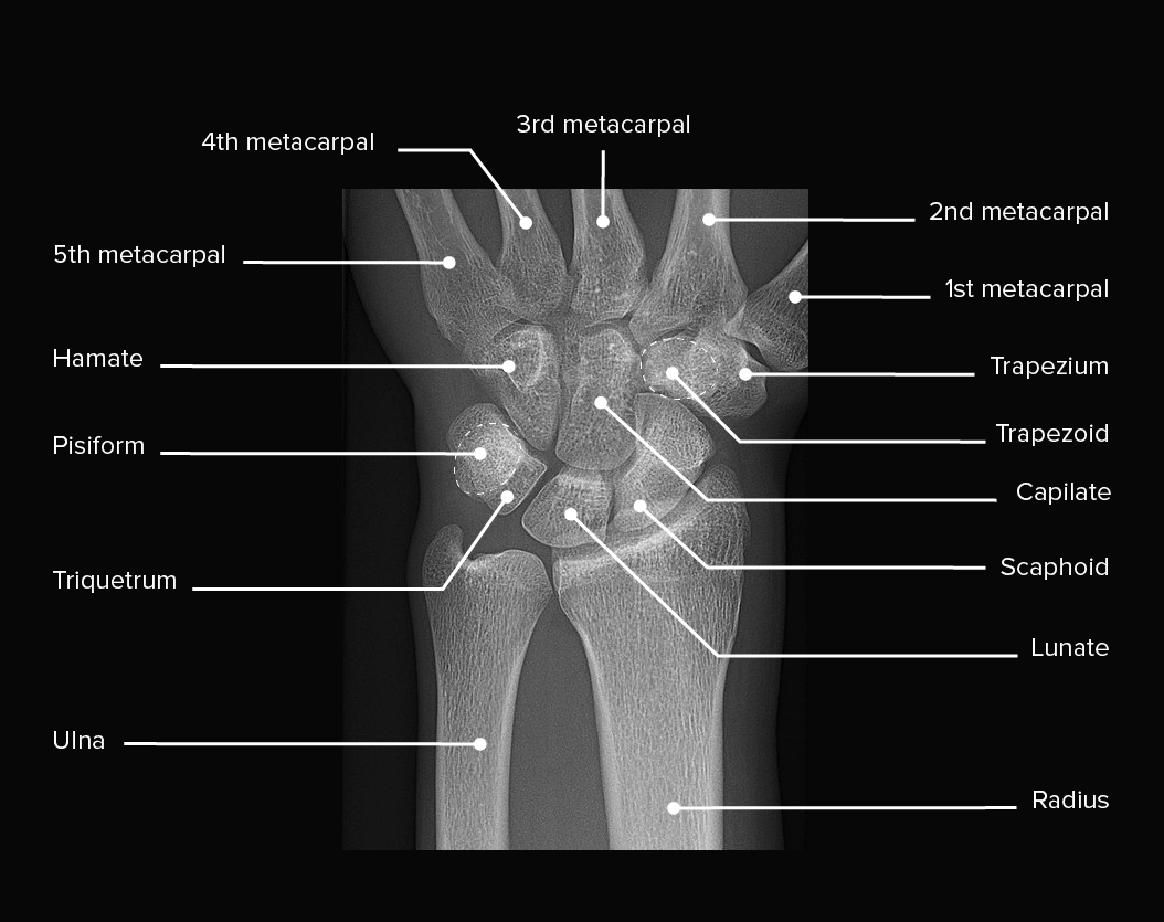
Wrist Radiographic Anatomy wikiRadiography
A wrist X-ray can help find the cause of common signs and symptoms such as pain, tenderness, swelling, or show deformities of the wrist joint. It can also de.
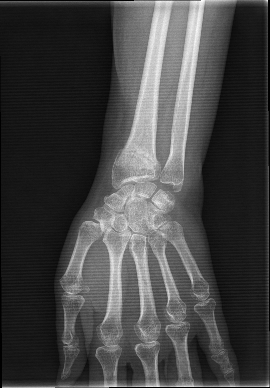
AP Wrist XRay
Anatomical structures of the upper limb using plain X-Rays. On "Anatomical parts" you can choose between two types of labels: bones and joints. On "Series" you can directly access the radiological images of the pectoral girdle, shoulder, arm, elbow, forearm, wrist, hand and fingers. All of the structures were labeled using the.

Lateral radiograph of the right wrist The BMJ
Introduction The wrist is one of the most complex joints in the human body, allowing for stability during movement in all three cardinal planes of the human body. Categorically considered a hinge joint like the elbow, the wrist has additional planes of movement and rotation thanks to robust anatomy.
Wrist Xray Interpretation OSCE Guide Geeky Medics
The wrist joint is an articulation of the distal head of the radius, the articular disc that overlies the distal ulna, and the proximal carpal bones of the hand (scaphoid, lunate and triquetrum). The carpal bones are arranged in a convex formation, whereas the other articular surface is concave. The main movements of the wrist are flexion and.

Wrist Anatomy
Understanding the key components of a normal wrist X-ray image is vital for proper evaluation: 1. Bones: Wrist X-rays highlight the various bones in the wrist, such as the radius, ulna, carpal bones, and metacarpals. 2. Joints: The spaces between these bones, or joints, are also visible in the X-ray. This is where conditions like arthritis can.

Plain Xray of right wrist anteriorposterior view and lateral view.... Download Scientific
The basic principles about the wrist X-ray examination. The basic principles about the wrist X-ray examination. Home; About us; Modules;. Normal Anatomy. The wrist is made up of a number of complex joints.. The joint spaces between the carpal bones are the same everywhere; in adults 1-2 mm. Broadening of a joint space suggests traumatic.

Xray Image of Wrist Joint Front View of Normal Wrist Joint Stock Photo Image of fracture
The radiocarpal joint is an articulation between the distal radius and the proximal carpal row of the wrist. It is a major synovial joint of the wrist and is an example of a condyloid joint . Gross anatomy Location

Wrist joint (Radiocarpal joint) Medically
The radiocarpal joint is a synovial joint formed between the radius, its articular disc and three proximal carpal bones; the scaphoid, lunate and triquetral bones.

Wrist Joint Xray
The wrist is a complex joint that connects the hand to the forearm. It is formed by the two bones of the forearm — the radius and the ulna — and eight small carpal bones. The carpal bones are arranged in two rows at the base of the hand. There are four bones in each row. The bones of the wrist.
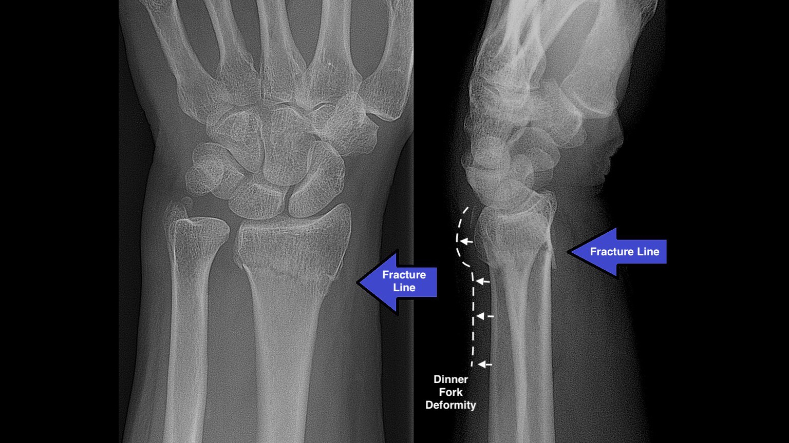
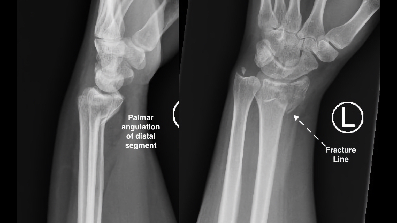
X Ray Wrist Joint Post Trauma Radiology Imaging
Bookshelf ID: NBK534779 PMID: 30521200. The wrist joint also referred to as the radiocarpal joint is a condyloid synovial joint of the distal upper limb that connects and serves as a transition point between the forearm and hand. A condyloid joint is a modified ball and socket joint that allows for flexion, extension, abduction, and adduction.
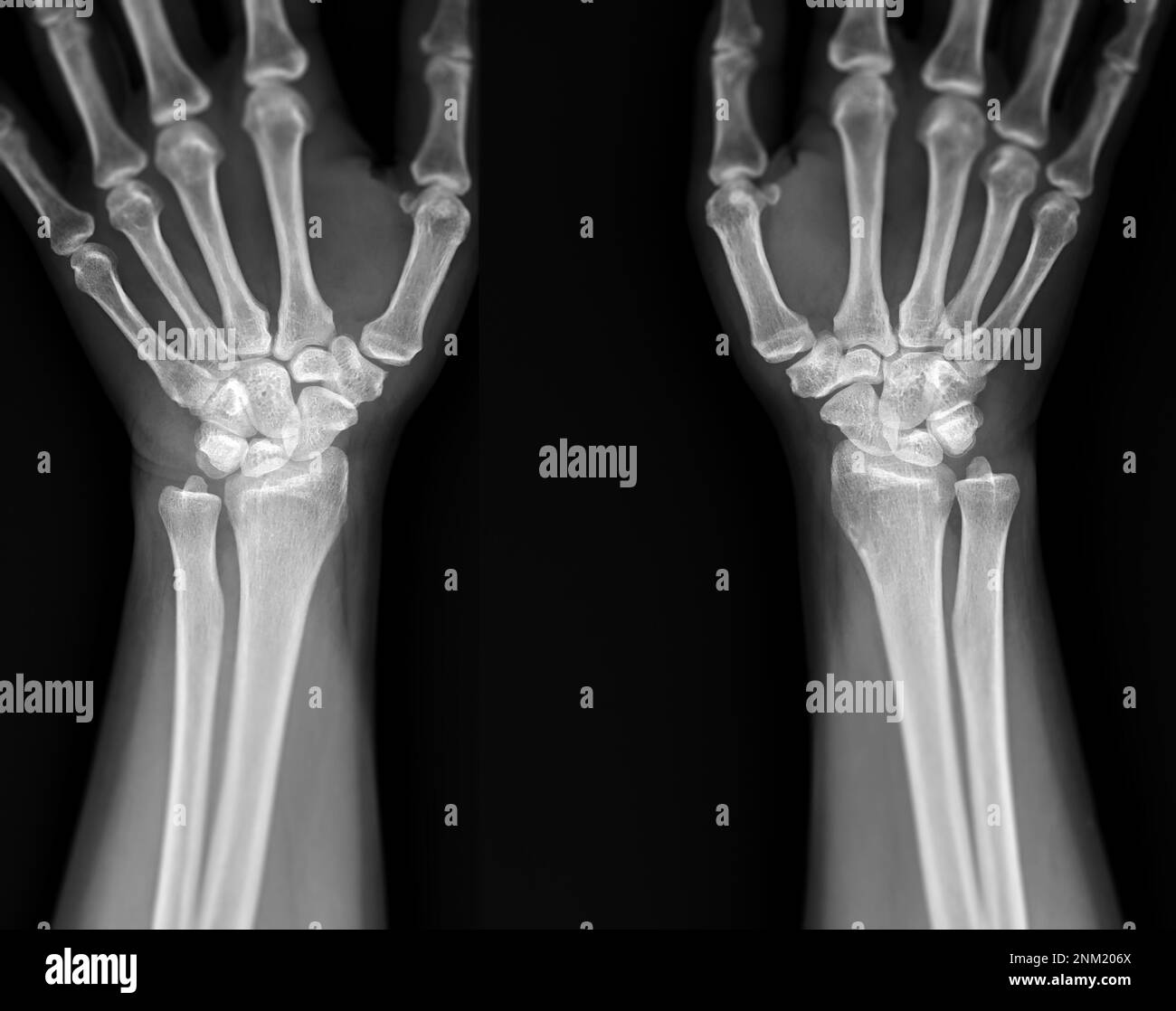
Xray image of wrist joint front view of normal wrist joint Stock Photo Alamy
A recommended systematic checklist for reviewing musculoskeletal exams is soft tissue areas, cortical margins, trabecular patterns, bony alignment, joint congruency, and review areas. Review the entire radiograph, regardless of perceived difficulty.
Wrist Xray Interpretation OSCE Guide Geeky Medics
Wrist Joint: Anatomy. The wrist connects the forearm to the hand. It consists of 8 carpal bones, multiple joints, and various supporting ligaments, as well as the distal bones of the forearm and the proximal portion of the 5 metacarpal bones of the hand. The wrist is crucial for the functioning of the upper limb, and it provides stability while.
Wrist Xray Interpretation OSCE Guide Geeky Medics
Wrist trauma is a common presentation to the emergency department and X-ray is typically the first-line investigation used to identify bony injuries. This guide provides a step-by-step approach to interpreting wrist X-rays and includes examples of the key pathology you may come across. Anatomy